Classics in the History of Psychology
An internet resource developed by
Christopher D. Green
York University, Toronto, Ontario
ISSN 1492-3173
(Return to index)
[Classics Editor's note: There are widespread inconsistencies in the use of punctuation, abbreviation, italics, and even spelling in the original publication of Sanford's "Course." These idiosyncrasies have been reproduced here as accurately as possible. The use of "[sic]" has been confined only to instances that might otherwise lead to confusion.]
By Edmund C Sanford (1891-1892)·
First published in American Journal of Psychology, 4, 141-155, 303-322, 474-490.
Posted June 2000
By Edmund C. Sanford (1892)·
(Third Paper [pp. 474-490].)
V. -- VISION.
THE MECHANISM OF THE EYE, AND VISION IN GENERAL.
Apparatus. Many oft he experiments of this section can be performed with very simple apparatus, made on the spot. The following materials will be needed : Pins, cards, corks, a candle, a couple of postage-stamps, a watch glass, pieces or colored glass, black and white card-board (not shiny), colored papers, a light wooden rod. Four inches square is a convenient size for the glass, of which two pieces should be cobalt blue, one red. Any colored papers will serve; those made for artificial flowers are easy to get in large variety of tints. A fine series of papers in Helmholtzian colors is sold by R. Jung, Heidelberg. In addition to these supplies there is need of a double convex lens short focus, two inches or more in diameter; an ordinary burning or reading glass would do, though those mounted on an adjustable stand, costing $2.50 and upward from the physical instrument dealers, are more convenient; also a concave spectacle lane.
For Ex. 99 a pink-eyed rabbit and a little modeling clay are necessary. An instrument for facilating[sic] Ex. 103 (a Phakoscope) can be had from Jung for 25 marks; a more elaborate instrument of the same name is quoted by the Cambridge Scientific Instrument Co., St. Tibb's Row, Cambridge, England, for £8-8.
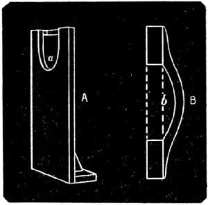
For Ex. 109 and other experiments a firm head rest of some sort is required. For most purposes one like that shown in the cut will answer well enough and can easily be made. Fig. A shows a board about 20 in. high and l2 in. wide with a U-shaped opening cut in the top to receive the lace, the chin resting at a. Fig. B, the top view, shows the cross piece against which the forehead rests at b. The whole when in use is clamped to the edge or the table. When a complete immobility of the head is desired it is best secured by providing a thin board cut out so that it can be put into the mouth and taken between the jaws. If the parts open which the teeth rest are covered with sealing wax and are bitten upon while the wax is still soft, not only is a firm support for the head seemed, but the head can be returned again exactly to its former position after an interval, if desired. Such a mouth board could [p. 475] easily be added to the support shown in the out. For pictures of such mouth boards cf. Hermann, Handbuch der Physiol. III, pt. 2, pp. 440, 473 and 478, also Helmholtz, Optique physiologique, p. 665 (p. 517 of the first edition) Aubert, Physiologische Optik, p. 647.
For Ex. 110 make a saturated solution of chrome-alum in water, filter and put into a flat-sided clear glass bottle. Dilute, if necessary, till the yellow spot can be observed as described in the experiment.
Ex. 115 requires a pair of electrodes and a battery. The electrodes can be made by soldering connecting wires to plates or brass or zinc, two and a half inches wide by three long and covering them with cloth. Some kind of a key for opening and closing the circuit and a commutator for changing the direction of the current are helpful, though not essential. Any battery giving a sufficiently strong current will do; one of four cells of the "gonda" pattern has proved sufficient for demonstrative purposes, and every much weaker one will serve for showing the flash from electrical stimulation.
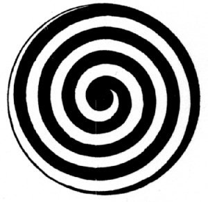
Ex. 119, involves a rotation apparatus of some kind and a disk traced with a spiral as in the cut. Any rotation apparatus will do, but if the laboratory is supplied with batteries one of the small electric motors now to be had at a very low price is easily adaptable for use and is extremely convenient.[1] A Porter motor, retailing at $3.00, has been used with success In this laboratory. It is well to have the disk large, a foot in diameter, and the line of the spiral thick, three eighths of an inch across, and a good black.
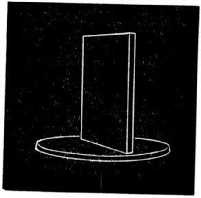
In a number of experiments black or white screens are to be used. A simple piece of black or white card-board will generally answer but sometimes the more permanent form indicated in the cut is convenient. It consists of an upright board 18 inches high, seven eights[sic] of an inch thick and 12 Inches wide, firmly fixed on a wooden base. In the cut the base in made too large. One side of the upright is covered with black card-board (or painted a dull black), the other with white card-board.
Of models helpful in understanding the mechanism and functions of the eye there are a number. Anatomical models are quoted, among others, by Jung (12 marks); by Kny & Co., 17 Park Place, New York, ($5.00 to $28.00); by Queen & Co., 924 Chestnut St., Philadelphia (Auzoux models, $19.00 and $20.00 without duty). Of physiological models, the best for accommodation and the like is Kühne's optical [p. 476] eye made by Jung, at 65 marks, by the Cambridge Scientific Instrument Co., at £7. The action of the muscles and the behavior or the eye in motion is illustrated by the Ophthalmotrope, described with cut by Helmholtz, Optique physiologique p. 678, (p. 527 in the German edition).This instrument is to be had of Jung, at 25 marks. Of the Cambridge Scientific Instrument Co., for £10; and of other dealers also. Another instrument for the same purpose, called the Blemmatotrope is described by Hermann in Pflüger's Archiv, VIII, 1873, p 305. The motions of the eye and their effect on the retinal image, such especially as those mentioned in Ex. 123, are finely shown by the Phenophthalmotrope of Donders, described in v. Graefe's Archiv für Ophthalmologie, Bd. XVI, 1870, and Bold by Jung at 30 marks. An improved form of the instrument is to be had of D.B. Kagenaar, Rijks-Universiteit, Utrecht, at 40 guilders. Suggestions for simple illustrative apparatus will be found with the description of the experiments.
Standards and rods with clamps and universal joints, thought not distinctively for visual experiments, are by far the most important of the general conveniences of a laboratory. They enter into the setting up of very many experiments and a liberal share of even a small appropriation may well be invested in them. Ordinary clamps can be bought in all sizes at the hardware stores at prices from ten cents upward. The standards and couplers to be had from the chemical and physical instrument dealers are made for another purpose and are not very satisfactory in the psychological laboratory. Those made for physiologists and photographers are better. Wilhelm Petzoldt, Bairische Str. 13, Leipsig[sic], makes a considerable variety of which the following have been found useful in the physiological and psychological laboratories of Clark University. Standards: simple tripods with interchangeable rods of 9 and 13 mm. diameter, 6.50 marks, and large tripods with leveling screws In two of the feet and carrying two of the above mentioned rods, at the same time, 16 marks. Table-clamps, which screw on to the edge of the table and are bored to receive the rode, thus taking the place of tripods: two kinds, one bored for the 9 mm. rods, but having only a vertical hole, 2.75 marks; the other bored for l3 mm. rods having both horizontal and vertical holes, 3.50 marks. Couplers to fit both sizes of rods: those for the 13 mm. rod (of iron) and connecting the rods only at right angles, 2 marks, those for the 9 mm, rods (of brass) and connecting the rods either at right angles or parallel 2.75 marks. Petzoldt also makes smell clamps of various sizes, like those furnished with the chemical sets, mounted upon the 9 mm. rods, at 3 marks. The advantage of these rods and couples, is that they fit nicely and can be set up so as not to wobble. By using several rods and couplers a universal motion can be secured, but not so conveniently, as by the ball-joint clamps and swivel couplers made for photographers' use by Otis C. White, of Worcester Mass. These allow extreme freedom or movement, and when fastened do not slip nor wobble. The ball joints are made to clamp on the edge of the table or to screw upon the end of rods. The first can be had in great variety of sizes, a convenient one fitting half inch rode costing $1.25. The swivel couplers allow the coupling of the rode in any position relative to each other, those of size to connect half inch and quarter inch rods costing 50 cents. Rods of various diameter and length may also be had with the ball-joints and swivel clamps. In purchasing for a laboratory from several makers it would be well to fix upon standard sizes for rods and fittings so that all may be interchangeable; and also to fix upon a standard size and number of threads to the inch for all screws cut upon the rods so that any clamps, pulleys or other small pieces of apparatus, made to screw upon one, [p. 477]
On Vision in general cf. Helmholtz, Handbuch der physiologischen Optik. (The second German edition has reached page 400: the latest complete edition is the French translation. Optique Physiologique, Paris. 1867). Aubert. Grundzüge der physiologischen Optik, Leipzig, 1876. (a portion of Graefe and Saemisch's Handhuch der ges. Augenheilkunde). Le Conte, Sight, New York. 1881. Beaunis, Nouveaux Éléments de Physiologie Humaine. Paris. 1888, (Beaunis, like Helmholtz, gives bibliographies). Wundt. Physiologische Psychologie. II, 82-209. Hermann's Handbnch der Physiologie. Bd. III, Th. 1, Leipzig. 1879.
The references following the experiments below are made chiefly to Helmholtz, the pages of the new German edition, the French edition, and, in parenthesis following the latter, of the first German edition being given, but the experiments of this section are more or less fully discussed In almost all of the works just mentioned and in many others besides.
99. The retinal image. The mechanisms of the eye accomplish two things: the projection of a well defined image on the retina; and the ready shifting of the eye so as to bring successive portions of the image into the best position for vision. The retinal image is readily seen in the unpigmented eye of a pink eyed rabbit. Chloroform the rabbit, remove the eyes and mount them in clay for readier handling. Make a thick ring of clay with an internal diameter a little greater than that of the comes of the rabbit's eye, place the eye comes downward in the ring and lay a similar ring upon it to keep it in place. It can now be handled easily and turned in any direction. Turn it toward the window and from behind observe the inverted image on the retina. Bring the hand into range and move it to and fro observe that the image of distant objects is more distinct than that of the hand. If convex and concave lenses are at hand (spectacle lenses will answer) bring them before the eye and observe that the effect upon the retinal image is similar to that seen subjectively when they are held before the observer's own eye. Reverse the eye, holding it retina side toward the window, and observe the radiating and circular fibres of the iris. The eye must be fresh, for if long removed it loses its transparency.
100. Accommodation. The sharpness of the retinal image depends on the adjustment or the crystaline lens, which must be such as to focus the light from the object under regard upon the retina. The lens must be thicker and rounder for near objects, thinner and flatter for more distant ones. These adaptations of the eye are known as Accommodation. The changes in the clearness of the retinal image are easy to observe subjectively. Hold up a pin or other small object six or eight inches away from the eyes. Close one eye and look at the pin with the other. The outline of the pin is sharp, but the outlines of things on the other side of the room behind it are blurred. Look at these and the outline of the pin becomes blurred. Notice the feeling or greater strain when looking at the nearer object. The experiment is somewhat more striking when the nearer object is a piece of veiling or wire gauze and the farther a printed page.
On this and the next two experiments cf. Helmholtz, Physiologische Optik, 2nd Ed. pp. 112-118, French ed. pp. 119 (90)-126 (96).
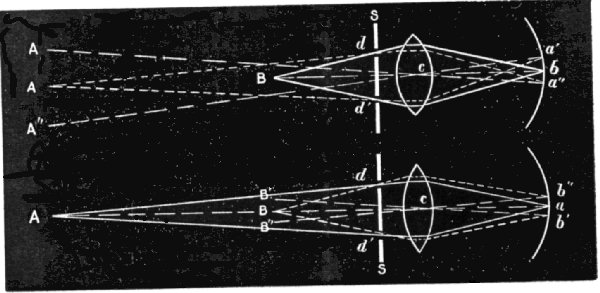
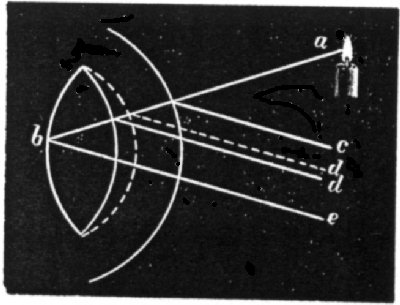
101. Accommodation. Scheiner's experiment. a. Pierce a card with two fine holes separated by a less distance than the diameter of the pupil, say, a sixteenth of an inch. Set up two pine in corks distant respectively eight and twenty inches in the line of sight; close one eye and holding the card close before the other with the holes in the same horizontal line look at the nearer pin; the farther pin will appear double; look again at the nearer pin and while looking cover one of the holes with another card; one of the images of the farther pin will disappear, the left when the left hole is covered, and the right when the right is covered. Look at the farther pin or beyond it and repeat the covering, covering the left hole now destroys the right image of the nearer pin and covering the right destroys the left. Why this should be so will be clear from the diagrams below. The upper diagram illustrates [p. 478] the course of the rays of light when the eye is accommodated for the nearer pin; the lower diagram when it is accommodated for the farther pin. A and B represent the pins; S and S the pierced screen; d and d' the holes in the screen; c and c the lens; a'ba" and b"ab' the retina; A', A", B', and B", the positions of the double images; the solid lines the course of the rays from the pin accommodated for; the dotted lines the course of the rays from the other pin; the lines of dashes the lines of direction, i.e., those giving the direction in which the images appear to the observer. In the upper diagram the rays from B are focused to a single retinal image at b, while those from A, being less divergent at first, are brought to a focus nearer the lens, cross over and meet the retina at a' and a", and since each hole in the screen suffices to produce a retinal image, cause the pin to appear double, and its two images are referred outward as usual with retinal images along the lines of direction, (which cross a little forward of the back surface of the lens, in the crossing point of the lines of direction), the right retinal image corresponding with the left of the double images and vice versa. If now the right hole in the screen be closed the left retinal image and the right double image disappear. The case of accommodation for the farther pin will be clear from the lower diagram, if attention is given to the dotted and dashed lines. It will also be easy to explain why moving the card when looking through a single pin hole causes apparent movements or the pin not accommodated for, and why in one case the movement seems to be with the card and in the other case against it. b. Stick the pins into the corks so that they shall extend horizontally, and examine them with the card so held as to bring the holes above one another. c. Arrange the holes thus: ![]() and observe that the triple image of the nearer pin (when the farther is fixated) has the reverse figure
and observe that the triple image of the nearer pin (when the farther is fixated) has the reverse figure ![]() Scheiner's experiment can easily be illustrated with a double convex lens and a pierced screen of suitable size.
Scheiner's experiment can easily be illustrated with a double convex lens and a pierced screen of suitable size.
102. The Range of accommodation. a. Find by trial the nearest point at which a pin seen, as In Ex. 101, can be seen single. This is the near point of accommodation. For the short-sighted a far point may also be found, beyond which double images reappear. b. Find how far apart in the line of sight two pins may be and yet both be seen single at one and the same time. Try with the nearer at 20 cm., at 50 cm., at 2 m. That portion of the line of sight, for points in which the same degree of [p. 479] accommodation is sufficient, is called the line of accommodation. The length of the line Increases rapidly as the distance of the nearer object from the eye increases.
Cf. Helmholtz, op. cit. G. 114, 119. Fr. 122 (93), 123 (97).
103. The Mechanism of accommodation. a. The change in the lens in accommodation is chiefly a bulging forward of its anterior surface. This may be observed as follows. Let the subject choose a far and a near point of fixation in exactly the same line or vision, class one eye and fix the other upon the tar point. Let the observer place himself so that he sees the eye or the subject in profile with about half the pupil showing. Let the subject change his fixation at request, from the far to the near point, being careful to avoid any sidewise motion of the eye. The observer will then notice that more of the pupil shows and that the farther side of the his seems narrower. This change is due to the bulging forward of the front of the lane. If the change were due to accidental turning of the eye toward the observer the farther edge or the iris should appear wider instead of narrower. b. Purkinje-Sanson images. The changes in the curvature of the lens may also be observed by means of the images reflected from its front or back surfaces and from the front of the cornea. Operate in a darkened room or at night. Let the subject choose far and near fixation points as before. Let the observer bring a candle near the eye of the subject at a level with it and a little to one side and place his own eye in a position symmetrical to the candle on the other side of the subject's line of eight. Careful examination will show three reflected images of the flame; one on the side of the pupil next the light, easily recognizable, bright and erect, reflected from the surface of the cornea; a second nearer the centre of the pupil and apparently the farthest back of the three, erect like the first, but very indistinct, (more like a light cloud than an image), reflected from the anterior surface of the lane; and a third, a mere point of light, near the side of the pupil farthest from the flame, inverted and reflected from the posterior surface of the lens. When the observer has found these three images the subject should fixate alternately the near and far points chosen. As he fixates the near point the middle image will grow smaller, advance and draw toward the corneal image; when he fixates the far point the image will enlarge, recede and move away from the corneal image. The following diagram after Aubert illustrates the movement of the middle image; the full lines indicate the positions of the comes and lens and the course or the rays of light when the eye is accommodated for the far point; the dotted lines indicate the anterior surface of the lens and the direction or the ray reflected from Its surface when the eye is accommodated for the near point. Three images similar to those in question can be observed on a watch glass and a double convex lens herd In the relation of the comes and crystaline.
Cf. Helmholtz, op. cit. G. 131-141. especially 131-134. Fr. 142 (104)-154 (112), especially 142 (104)-146 (101). Aubert, Physiologische Optik. 444.
104. Chromatic aberration. Of the various defects of the eye as an optical instrument only one will be mentioned here, namely, chromatic aberration, and that because it has been supposed to offer a possible means of inferring the relative distance of objects from the eye. The [p. 480] different colored rays of light are not equally retracted by the lens, the violet most, the red least, and the other colors In order between. The point at which parallel violet rays are brought to a focus is therefore nearer the lens than the point for red; and in order that the same degree of accommodation may serve to show a red lighted object and a violet lighted object at the same time and both with full distinctness, the red must be somewhat farther away. a. The aberration can easily be observed by looking at a small gas or candle flame through a piece of cobalt blue glass which transmits light from the two ends of the spectrum chiefly. Hold the glass eight or ten inches before the face and fixate some point on it the flame will appear pinkish with a blue border. Fixate some point considerably beyond the flame; the flame is now bluish and the border is a fine red line. b. Look at the edge of the window frame next the pane, and bring a card before the eye so about half the pupil is covered; if the card has been brought up from the frame aide, the frame will be bordered with yellow; if from the pane side, with blue. In ordinary vision these fringes do not appear, because the colors overlap one another and produce a practically colorfess [sic] mixture. c.v. Bezold's experiment. Something similar may be observed, on regarding the parallel lines of the left figure under Ex. 111 with imperfect accommodation.
Cf. Helmholtz. op. cit. G. 156-164; Fr. 172 (125)-119 (131). Beaunis, Nouveaux éléments de physiologie humaine. II. 506. v. Bezold, v. Graefe's Archiv F. Ophthalm., XIV, Heft 2, 1-29.
105. Accompanyments or accommodation. a. Notice that as the subject in Ex. 103 accomodates [sic] for a near point, his pupil grows smaller, and as he accommodates for a far point, grows larger. Cf. Also Ex. l06, b. Degrees of accommodation suitable for objects at different distances are habitually associated with the amounts of convergence of the lines of sight necessary to fix the eyes upon such objects, and a little practice is necessary before the convergence and accommodation can be dissociated. Place a couple of postage stamps six inches apart on the table and look at them from a distance of twelve or fifteen inches with crossed eyes so that the left eye looks at the right stamp and the right eye at the left stamp; the lines of sight now cross only a few inches from the eyes and the accommodation is for that distance and not for the true distance of the stamps, as is betrayed by the blurring of their images. Holding a pencil at the crossing point of the lines of sight is helpful in first attempts at crossed vision.
Cf. Helmholtz, op. cit. G. 130. Fr. 142 (104).
106. Entoptic phenomena: Muscae volitantes, etc. Fix a lens of short focus at some distance from a bright gas or candle flame. a. Set up in the focus of the lens a card pierced with a very fine hole, bring the eye close to the hole and look toward the light; the eye should be far enough from the hole to prevent the edge of the lens from being seen; the rays of light that now reach the eye are divergent and the crystaline lens does not bring them to a focus on the retina, but only refracts them to such a degree that they traverse the eye nearly parallel and thus in suitable condition for casting sharp shadows upon the retina or objects on or in the eye. The lens will appear full of light, and in it will be seen a variety of shadings, blotches and specks, single or in strings, the outward projection of the shadows just mentioned. The figures in this luminous field will vary from person to person, even from eye to eye, but in almost every eye some will be found that move and some that remain fixed and only move with the eye. Of the moving figures some are due to particles and viscous fluids on the surface of the eye; they seem to move downward and are changed by winking. Notice for example the horizontal bands that follow a slow dropping and raising of the upper [p. 481] lid. Others, the muscae volitantes are frequently noticed without any apparatus, they appear as bright irregular threads, strings of beads, or groups or points, or single minute circles with light centres. They seem to move downward in the field and consequently actually move upward in the vitrious humor where they are found. Of the permanent ones, some are due to irregularities of structure or small bodies in the lens and its capsule (spots with dark or bright centres, bright irregular lines, or dark radiating lines corresponding probably to the radial structure of the lens); others of a relatively permanent character can be produced on the cornea by continued rubbing or pressure on the eyeball. b. The round spot of light in which these things are seen represents the pupil, and the dark ground around it the shadow or the iris. Notice the change in the size of the spot of light, as the eye is accommodated for different distances (cf. Ex. 105), and as the other eye is exposed to, or covered from, the light. The change begins in about halt a second. It shows the close connection of the iris mechanisms or the two eyes and is typical of the way in which the two eyes co-operate as parts of a single visual machine. Some of these entoptic observations may be made with a pierced card alone, or simply by looking directly at a broad expanse of clear sky with out any apparatus at all.
Cf. Helmholtz, op. cit. G. 184-192 and Tafel I. which represents the appearance of several of the entoptic objects; Fr. 204 (149)-214 (156) and Pl. V; also pp. 548 (419)-558 (427).
107. Retinal blood-vessels, Purkinje's vessel figures. a. Concentrate a strong light, (preferably in a dark room) or even direct sunlight, with a double convex lens of short focus on the sclerotic in the outer corner of the eye of the subject, requesting him to turn the eye toward the nose and giving him a dark background to look toward. Make the spot of light on the sclerotic as small and sharp as possible and give to the lens a gentle to and fro or circular motion, and after a little the subject cannot fail to see upon the field which the light makes reddish yellow the dark branching figure of the shadows of the retinal vessels. Notice that the area directly fixated, is partially surrounded, but not crossed by the vessels. In this lies the yellow spot (macula lutea) or area of clearest vision of the retina, not, however, to be observed in this experiment. The centre from which the vessels radiate lies in the point or entrance of the optic nerve. In this form of the experiment the light radiates in all directions within the eye from the illuminated point of the sclerotic. b. Somewhat the same kind of an image of the vessels is to be secured by moving a candle about near the eye, below it and a little to one side. In this experiment some indication of the region of the yellow spot is to be seen. In this form of the experiment the light enters by the pupil, forms an image on a part of the retina somewhat remote from the centre and this retinal image is the source of light by which the vessel shadows are cast. c. Look through a pin hole in a card directly at the clear sky or any other strongly illuminated even surface or at a broad gas flame. Give the card a rather rapid circular motion and the finer retinal vessels in the region of the yellow spot will readily be seen, among them also a small colored or slightly tinted spot (best seen perhaps by gas light) representing the macula, and in its centre a shadowy dot (representing the fovea or point of clearest vision) which appears to rotate when the motion of the card is circular. If the card is moved horizontally the vertical vessels alone appear; it vertically, the horizontal vessels. Notice also the granular appearance of the macula; the granulations have been supposed to represent the visual cones of that region. The finer retinal vessels can also be seen when looking at the vacant field or a compound microscope, it the eye is moved about rapidly. In all of these cases it is important that the shadows be kept moving; it they stand still, they are lost. The explanation [p. 482] is partly physiological, the portions of the retina on which the shadows rest soon gain in sensitiveness enough to compensate for the less light received, and partly psychological, moving objects in general being more readily attended to, and those whose images rest continously [sic] on the retina without motion being particularly subject to neglect. Once having become acquainted with the appearance of these vessel figures it is often possible to see traces of them without any apparatus. Parts of them, with something of the projection of the yellow spot, map sometimes be seen for an instant as dark figures on the diffusely lighted wells and ceiling or as light figures on the dark field or the closed eyes when the eyes are opened and closed after a glance at the window on first waking in the morning, or in blue when looking at the snow and winking on a bright morning, or projected on the sky and keeping time with the pulse after a rapid walk up hill.
Helmholtz op. cit. G. 192-198. Fr. 214 (156)-211 (161).
108. Retinal circulation. Look steadily through two or three thicknesses of blue glass at the clear sky or a bright cloud, and observe a large number of what seem to be bright points darting hither and thither like bees in a swarm or rapidly blown snow-flakes. Careful observation will also establish that the bright points are followed by darker shadowy ones. Pick outs speck on the window to serve as a fixation point, look at it steadily and observe that while the movements or the points seem irregular the same lines are retraced by them from time to time. When several of their courses have been accurately observed, repeat the experiment for demonstrating the finer retinal vessels (Ex. 107 c.) and notice that fine vessels are found which correspond to the courses which the points seem to follow. These flying points can be seen without the glass by a steady gaze at an evenly lighted bright surface, and some times a rhythmic acceleration of their movement will be found, corresponding to the pulse. Helmholtz explains the phenomenon as due to the temporary clogging of fine capillary vessels by large blood corpuscles. The bright lines (the apparent tracks of bright points) are really the relatively empty capillary tubes ahead or the corpuscles, which, after an instant, are driven onward by others crowding behind and in turn give the shadow that apparently follows the bright points.
Cf. Helmholtz, op. cit. G.198; Fr. 221 (837), 555(425), Rood, American Journal of Science, 2d Series, XXX, 1860, 264-265, 385·386.
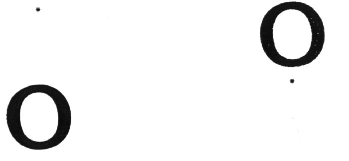
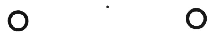
109. The Blind-spot. Mariotte's experiment. The point of entrance or the optic nerve is unprovided with visual end-organs and is irresponsive to light. a. This insensitiveness is easily demonstrated with the diagrams below. Close the left eye and keeping the right fixed on the asterisk in the upper diagram move it backward and forward till a point is found where the black oval disappears. For the blind spot of the [p. 483] left eye use the second diagram. The blind spot may be demonstrated simultaneously in both eyes by the use of a figure like that below enlarged a couple of times. The experimenter should look at the asterisk while he holds a sheet of paper in the median plane of his head, to prevent each eye from seeing the other's part of the diagram.
b. To draw the projection of the blind-spot, arrange the head support described above, piece opposite the face at a distance of about 18-inches, a vertical sheet or white paper and put a dot on it for a fixation point. Fasten upon the end of a light rod a bit of black paper about 2 mm. square or blacken the end of the rod with ink. Bring the face into position, close one eye, and fix the other upon the dot. Move the rod slowly so as to bring the little square over the part or the white paper corresponding to the blind spot, dotting on the paper the points where the square disappears or reappears. Repeat at various points till the outline of the projection of he blind spot is complete. If the mapping is carefully carried out, the map will probably show the points of departure of the large blood vessel, that enter with the nerve.
Helmholtz, op. cit. G. 250-254, Fr. 284 (210)-288 (214).
110. The yellow spot, macula lutea. The projection of the yellow spot in the visual field can be made visible in several ways. Two have already been mentioned in Ex. 107; others are as follows. a. Close the eyes for a few seconds and then look with one or them through a flat sided bottle of chrome alum solution at a brightly lighted surface (not yellow) or the clear sky. In the blue green solution a rose colored spot will be seen which corresponds to the yellow spot. The light that comes through the chrome alum solution is chiefly a mixture of red and green and blue. The pigment or the yellow spot absorbs a portion of the blue and green and transmits the rest, which makes a rose colored mixture, to the visual organs behind it. b. The region of the yellow spot may be seen as an area of somewhat deeper shade when the eye looks at on evenly lighted surface like the ceiling, and the illumination is made intermittent by moving the spread fingers to and fro between the eye and the ceiling.
Cf. Helmholtz, op. cit. Fr. 548 (419)-551 (421). On a. cf. Maxwell, On Color-vision at different points of the Retina, Report of the British Assoc., 1870; or Vol. II, pp. 230-232 of Maxwell's Scientific Papers. Cambridge, 1890.
111. Visual cones in the fovea. Bergmann's experiment. Place the left hand diagram in a good light and look at it from a distance of a yard and a half or two yards. Observe the apparent bending and beading of the lines. This is supposed to be due to the mosaic arrangement [p. 484] or the visual cones. The cones that are touched by the image of one of the white lines are stimulated in proportion se are more or less touched. Those that are much stimulated furnish the sensation of the white line and its irregularities, those that are little stimulated join with those that are not touched at all to give the image or the black line and its irregularities. This is schematically represented in the right hand cut.
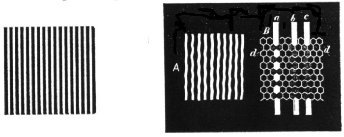
Cf. Helmholtz, op. cit., G. 257-258, Fr. 293 (217)-294 (218). Bergmann, Zeitschrift für rat. Med., (3), II, 88.
112. Acuteness of vision, minimum visibile, and size of the cones in the fovea. Place the parallel line diagram used in the last experiment in a good light and walk backward from it till the lines can just no longer be distinguished as separate. If the experimenter's eyes are not normal he should use glasses that fit his eyes for distinct vision at the distance required. Measure the distance between the eye and the diagram and calculate the angle whose apex lies in the crossing point of the lines of direction (about 7 mm. back of the cornea and 16 mm. in front of the retina) and whose base is the distance from the middle of one line of the diagram to the middle of the next; in this diagram 1.58 mm. This angle measures the least visible extent when discrimination is involved; the least luminous extent that can still impress the retina is far smaller, as witness the visibility or the stars. On the supposition that if the sensations of two cones are to be separable they must be separated by an unstimulated, or at least by a less stimulated, cone, it has generally been considered that the cones could not subtend a greater angle than that found in this experiment, 60"-90" representing 0.004-0.006 mm. on the retina, and this agrees well with microscopical measurements. But as Helmholtz notices(Phys. Opt. 2nd ed. p. 260) this experiment does no more than prove that there are on the retina rows of sensitive elements the middle lines of which are separated by the angular distance found in the experiment. The elements themselves, if properly arranged may be somewhat larger. Calculation of the number of such elements in a sq. mm. of the retina based on this view of the experiment agrees well in the case of Helmholtz's own determination with the result of microscopical counting. b. The discriminative power or the retina falls off rapidly in all directions from the fovea, more rapidly above and below than in a horizontal direction. Arrange a head rest and perpendicular plane as in Ex. 109 b. Place upon the end of the rod used in that experiment a card on which have been made two black dots 2 mm. in diameter and 4 mm. from centre to centre. Move the card horizontally toward the fixation point, beginning beyond the point at which the two dots can be distinguished and moving inward till they can just be distinguished. Measure the distance from the fixation point and repeat several times both to the right and left of the fixation point and above and below, holding the card so that both dots are in each case equally distant from the fixation point.
Helmholtz, op. cit., G. 255-264, Fr. 291 (215)-301 (223).
113. Mechanical stimulation of the retina. a. Phosphenes. Turn the open or closed eye as far as possible toward the nose and press on the eyelid at the outer corner with the finger or the tip of a pen holder. On the opposite side of the visual held will be seen a more or less complete circle of light surrounded by a narrow dark band, outside of which again is a narrow band of light. Notice the color of the light seen. Get the phosphenes by pressure at other points of the eye ball. b. Press the eye moderately with some large object, say the angle of the wrist when the hand is bent backward, and continue the pressure for a minute or two. Peculiar palpitating figures will be observed and [p. 485] strange color effects. The former Helmholtz compares to the tingling of a member that is "asleep." c. Standing before a window close the eyes and turn them sharply from side to side. As they reach the extreme position in either direction observe immediately In front of the face a sudden blue spot surrounded by a yellow band. A second fainter spot farther from the centre in the direction of motion may also be seen. The yellow ring is due to the stimulation of the portion of the retina in the region of the blind spot in the eye that turns inward. The blue spot represent the blind spot in the same eye. Cf. Explanation in the latter part of Ex. 115.
Helmholtz, op. cit. G. 235-259. Fr. 266 (196)-270 (200). Le Conte, American Journal of Psychology, III. 1889-90, 364-366.
114. Idio-retinal light, light chaos, light duet. Close and cover the eyes so as to exclude all light, or experiment in a perfectly dark room. Let the after effects of objective light fade away and then watch the shifting light clouds of retinal light. The cause of the retinal light is not altogether clear, but it is supposed to be a chemical action of the blood on the nervous portion of the visual apparatus. Aubert estimates its brightness at about half the brightness of a sheet of paper illuminated by the planet Venus when at its brightest. b. When awake in the night time in a room that is almost perfectly dark (e.g. in which the form of the window and the large pieces of furniture cannot be made out) notice that the white clothing of the arms can be seen faintly as they are moved about, but not when they are still. In the last case the very faint light they reflect is not sufficient to make them distinguishable from clouds of idio-retinal light.
Cf. Helmholtz op. cit. G. 242-243, Fr. 274 (202)-275 (203). On b. cf. Helmholtz. Die Störung der Wahrnehmung kleinster Helligkeitsunterschiede, Zeitschrift für Psyhologie. I, 1890, 6-9.
115. Electrical stimulation of the visual apparatus. Moisten thoroughly with strong salt water both the electrodes and the portions of the skin to which they are to be applied. Place one of the electrodes on the forehead (or on the edge of the table and lay the forehead upon it), the other on the back of the neck; or, if the current is strong enough, hold it in the hand or lay it on the table and put the hand upon it. At each opening or closing of the circuit a bright flash will be seen, whether the eyes are closed or open. With the eyes closed and covered the effects of the continuous current may be observed. In this case it is well to apply the electrode slowly and carefully so as to avoid as much as possible the flash caused by the sudden closing of the circuit. When the positive electrode is on the forehead, the negative on the back of the neck a transient pale violet light will be seen distributed generally over the field and forming a smell bright spot at its centre. Sometimes traces of the blind spot appear. The violet light soon fades and on opening the circuit, there is a notable darkening of the held with a momentary view of the blind spots as bright disks. When the negative electrode is on the forehead, the: positive on the back of the neck, the phenomena are in general reversed, the darkening occuring [sic] on closing the circuit, the violet light on opening it. Helmholtz sums up these and other experiments as follows: "Constant electrical circulation through the retina from the cones toward the ganglion cells gives the sensation of darkness, circulation in the contrary direction gives the sensation of brightness." (Phys. Opt. 2nd ed. 247). That the blind spot should appear as a disk of different color from the rest of the held seems to be due to the fact that the sensitive parts of the retina immediately surrounding it are somewhat shielded from the electric current, and as usual their condition is attributed to the blind spot also. The experiment is not entirely a pleasant one, on account of the feeling [p. 486] which the current produces in the head, the electrical taste in the month and the reddening of the skin under the electrodes.
Cf. Helmholtz, op.·cit. G. 243-248, Fr. 275 (203)-281 (207).
116. After-images, accidental or consecutive images, After-images in which the relations of light and shade of the original object are preserved are called Positive After-images. Those in which these relations are reversed (as in a photographic negative) are called Negative After-images. Positive after-images are of changing colors, but most important to notice here are those of the color of the object (like colored), and of the complementary color (opposite colored). Negative after-images, so far as observed, are always opposite colored. All after-images, especially the positive, can best be observed in the morning when the eyes are well rested. a. Negative after-images: look steadily for a minute at a fixed point or the window, then at a white screen or an evenly lighted unfigured wall; the dark parts of the window will now appear light and vice versa. Get a lasting after-image and look at a corner of the room or at a chair or other object of uneven surface; notice how the image seems to at itself to the surface upon which it rests. After a little practice it is also possible at desire to see the image floating in the air instead of lying on the background. b. Look steadily at a bright colored object or some bits of colored paper, then at the screen; observe that the colors of the after-images are approximately complementary to the colors of the objects producing them. Negative after-images are some times very lasting and for that reason are those most frequently noticed in ordinary experience; they are a phenomenon of retinal fatigue. c. Positive after-images. Look for an instant (one-third of a second) at the window, then close and cover the eyes, or look at a dark surface; for a very short time an after-image like the original object in color and distribution of light and shade can be seen. The positive after-image is of short duration and is not so readily observed as the negative; it is a phenomenon of retinal inertia, or the prolongation of retinal excitation. d. Colored positive after-images. Look for an instant at a gas flame through a piece or red glass, then close the eyes and observe the red image; repeat the experiment continuing the fixation of the flame for half a minute; the resulting after-image will be bright as before but of the complementary color. e. Get an after-image of the window of not too great an intensity, and alternately project it on a sheet of white paper and the dark field of the closed and covered eyes; it will be found negative on the white back-ground and positive on the dark. f. Get a good after-image of the window and observe with closed and covered eyes the play of colors as the image fades. Try several times and observe that the order of succession is the same.
Cf. Helmholtz, op. cit. Fr. 446 (338), 471 (357)-500 (380). Wundt, Physiologische Psychologie, 3rd ed I,472-476.
117. Effect of eye-motions on after-images. Get a moderately strong after-image of the window; look at the wall and keep the eyes actively in motion; the image will be seen with difficulty while the eye is in motion; when the eye is brought to rest, however, it will soon appear. In general any visual stimulus that moves with the eye is less effective than one that does not.
Cf. Exner, Das Verschwinden der Nachbilder bei Augenbewegungen. Zeitschrift für Psychologie, I, 1890, 47-51.
118. The seat of the after-image. An after-image due to exclusive stimulation of a single eye may under proper conditions sometimes seem to be men with the other unstimulated eye. From this it has been inferred that the seat or after-images was central, not peripheral; that is, in the visual centres of the brain, not in the [p. 487] eye. The following experiments show, however, that the after-image is really seen with the eye first stimulated, and so render the hypothesis of a central location unnecessary. a. Look steadily for several seconds at a bit of red paper on a white ground, using only one eye, say the right, and keeping the other closed; when a strong after-image has been secured, remove the paper, close the right eye, open the left and again look steadily at the white ground; after a little the field will darken and the after-image will reappear. If the red does not produce a sufficiently lasting image, substitute for it a gas flame or some other bright object. That we have really to do, however, with the eye originally stimulated, (its present dark held being superposed upon the light one of the other eye) appears from the results of b and c. Get the after-image as before; then open both eyes and bring a bit of card-board before the eyes alternately; bringing it before the left eye rather brightens the image bringing it before the right dims or abolishes it; the image is therefore chiefly affected by what affects the right eye. c. Get the after-image again and close and cover both eyes; observe the color of the after-image as projected on the dark held; then open the left eye, letting the right eye remain closed and covered; the after-image will be seen, not in the color it has when the right eye is open and the image is projected in the light field, but in that which it has in the dark field of the closed eye.
Cf. Delabarre. On the seat of Optical After-Images, American Journal of Psychology, II. 1888-89, 326-328.
119. After-images of motion. Fasten upon the rotation apparatus a disk like that in the first cut on page 475. Then look at a page of print or into the face of a by-stander and notice the apparent shrinking (if the spiral has seemed to run outward) or swelling (if the spiral has seemed to run inward). Illusions of increase or decrease of distance sometimes accompany those of motion. These after-images of motion have been explained as due to unconscious persisting movements of the eyes. This is probably incorrect, for in the present case it would seem necessary that the eyes should move in all directions at the same time.[2]
Cf. Helmholtz, op. cit. Fr. 766 (603)-769(605). Bowditch and Hall, Optical Illusions of Motion, Journal of Psychology, III, 297-307. Mach, Bewegungsempfindungen, Leipzig, 1875, pp. 59-61. (see also pp. 61-65 for yet another kind of after-image) and Analyse der Empfindungen, Jena, 1886, pp. 65-67.
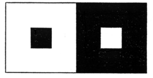
120. Irradiation. This term is used to designate the apparent enlargement of bright surfaces at the expense or adjacent dark surfaces. It is most strongly marked when the bright surface is intense and the accommodation is imperfect, but is not absent with perfect accommodation. Even with perfect accommodation, and much more so with imperfect accommodation, the line of juncture of a bright and dark surface is not really a sharp line but a narrow band of gray of which more than [p. 488] the proper amount is credited to the white, for reasons to be brought out in the section to follow on the Psychophysic Law. The following are some of the common cases of irradiation: a. Hold a ruler or a straight edged piece or black card-board close before a gas or candle flame so se to cover a portion of it, and notice that the flame seems to cut into the edge, and if there are differences in brightness the brightest parts cut in deepest. b. Notice that the white squares in the diagram below, when brought into a strong light, seem larger than the black, though they measure the same in size.
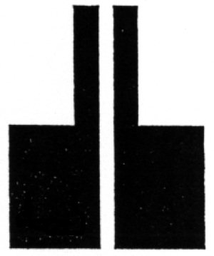
c. Irradiation of dark lines. A black line on a white surface (or a white line on a black surface) may some times be enlarged by the greater part or its gray fringe, because near the outer edge of the fringe the blackness (or for white lines, the whiteness) decreases very rapidly and so seems to make a boundary. Look at the accompanying diagram through a lens that will make accommodation very imperfect. The narrow black strips will appear larger for the reason just mentioned, while the lower black areas will be cut into as in the ordinary cases of irradiation, giving to the white stripe between the shape of a club with the handle uppermost. Helmholtz suggests with reason that these two phenomena, having quite different causes, should have different names, and the term "irradiation" be confined strictly to such enlargement of white surfaces as takes place with exact accommodation.
Cf. Helmholtz, op. cit., G. 394-400, Fr. 425 (321)-433 (327).
121. Reflex movement of the eye. The eye is a mooing as well as a seeing member and its motor functions are of great importance for psychology. Of the first importance is the constant reflex tendency of the eye to move In such a way as to bring any bright image lying on a peripheral part of the retina, or any to which attention is directed, into the area of dearest vision. Many evidences of this tendency will be found in the ordinary course of vision. By way of experiment, try to study attentively a musca volitans or a negative after-image that is just to one side of the direct line of sight. The apparent motion of the object measures the energy of the reflex.
122. Associated movements of the eyes. The two eyes form a single visual instrument and even when one eye is closed it follows to a considerable degree the movements of its open companion. a. Close one eye and, resting the finger-tip lightly on the lid, feel the motions of the eye as the other looks from point to point of the visual held. b. Get a monocular after-image as in Ex. 118 and when it has become apparently visible to the open eye, notice that it seems to accompany that eye as it takes one fixation point after another in the field of regard.
123. Motions of the eyes when the lines of sight are parallel, Donders's and Listing's laws. All motions of the eye can be interpreted as rotations of greater or less extent about one or more of three axes: a sagittal axis, corresponding nearly with the line of sight; a frontal axis, extending horizontally from right to left; and a vertical axis. All these intersect in the centre of rotation of the eye. Now it is easily conceivable that for any position of the line of sight, e.g. 15° to the right and 10° upward, there would be an infinite number of positions that the eye might assume by rotation about the line of sight itself. As a matter of fact, however, it does not assume an indefinite number of positions, but one and only one, no matter by what route the line of sight may have come to that point. This is the law of constant orientation [p. 489] or Donders's law. Listing's law goes further and asserts that the position is not only fixed, but is such as the eye would assume if the line of sight were moved from its primary position (approximately that in which the eye looks straight forward to the horizon) to the point in question without any rotation at all about the line of eight, but about a fixed axis standing perpendicular at the centre of rotation to both the primary and the new position of the line of sight. The advantage to vision of the constancy or orientation and the exclusion of rotation about the line of sight is considerable, especially in determining directions in the held of regard. The correctness of these laws is easy to demonstrate, a. Donders's law. Cut in a sheet of black cardboard two slits an eighth of an inch wide and six or eight inches long, crossing at right angles. Set the cardboard in the window or before some other brightly lighted surface. Arrange a head rest at some distance and when the head is in position, get a strong after-image of the cross, fixating its middle point. Then, without moving the head, turn the eyes to different parts of the walls and ceiling. The image will suffer various distortions from the different surfaces upon which it is projected, but each time the eye returns to the same point the image will lie as before. If the wall does not offer figures by which this can be shown, have an assistant mark the position of the image upon it. The after-image is of course fixed on the retina and can move only as the eye moves. b. Listing's law. Make over the cross used in a into an eight rayed star by cutting two other narrow slits across its centre. Arrange the card before a brightly lighted wall and parallel to it at a height a little less than that of the eyes when the head is in position. Draw lines or stretch threads on the wall that shall appear to continue the rays of the star upward and right and left, and downward if convenient. Fix the head rest directly before the star at a distance of five or six yards or more, adjust the head so that when the after-image of the star is carried along the horizontal or vertical line its corresponding ray will coincide exactly with the line. When this condition is fulfilled for both lines the eyes and lines of sight are in the primary position. When the primary position has been found, carry the after-image along the lines prolonging the other rays and observe that as before the after-image of the ray coincides with its line. This would be found true, for all except extreme positions, of all other rays, and shows that the eye does not in such motions rotate about the line of sight. c. In motions from other or secondary positions, however, there Is such a rotation. Turn the head somewhat to one side or tip it forward or backward from the primary position repeat b and notice that the lines of the after-image betray some rotation.
Cf. Helmholtz, op. cit., Fr. 601 (462)-610 (470), 621 (479) ff. Le Conte, Sight, pp. 164-177.
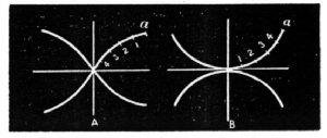
124. Actual movements of the eyes. Rapid motions of the eyes are not executed with mechanical exactness according to Listing's law, though it gives correctly the end position reached. The axis of rotation is not quite constant and the lines passed over by the point of sight are therefore not quite straight. This is easy to observe; as follows. In a dark room turn down the gas till it burns in a flame not more than 8 or 10 mm. high. Then using this as a point of departure in the primary position look suddenly from it to other points of fixation in various directions about it, and notice the shape of the long positive after-images that result from the motion of the image of the flame, over the retina. These will probably have the shape of the radii in the left hand figure below. The newest part of the after-image is that next the light, the oldest part is that next the fixation point, for example at a. If the points of the after-image curve are now interpreted in the order or time, it [p. 490] appears that the eye at first moved rather rapidly toward the right but rather slowly upward, while at last it moved rather slowly toward the right and rapidly upward. Plotting the curve accordingly we get the reverse curve shown in B which shows the true track of the fixation point. It is said that for some eyes the after-images, though curved, do not coincide with those figured in A.
Cf. Wundt. Beiträge zur Theorie der Sinneswahrnehmung, Leipsig[sic], 1862, pp. 139 ff. 202. Hermann's Handbuch der Physiol. III, Th. 1. 450-451.
125. Convergent movements of the eyes. When the lines of sight converge, the movements of the eye do not follow Listing's law. When the lines of sight converge in the primary position both eyes rotate outward; as the lines of sight are elevated, the convergence remaining the same, the outward rotation increases; as they are depressed, the rotation diminishes and finally becomes zero. On a sheet of cardboard draw a series of equi-distant parallel vertical lines one or two inches apart and eight or ten inches long, drawing the left half of the group in black ink, the right half in red. Cross both sets midway from top to bottom by a horizontal line, red in the red set and black in the black set. Fasten the cardboard fist upon a vertical support and arrange the head rest in front of it. The horizontal line of the diagram should be on a level with the eyes. a. Fasten a bit of wire vertically between the eyes and the diagram in such a way that it can be moved to and from the eyes. Bring the head into position and look at the wire, but give attention to the diagram. It will be seen that the red and black lines are not quite parallel and that they are less nearly so as the wire is brought nearer the face. The red lines (seen by the left eye) seem to incline a little toward the right and the black lines (seen by the right eye) toward the left. As the wire comes near and the convergence is great the horizontal lines will also show the rotation. This apparent rotation of the lines is not, as in the case of the after-images, a sign that the corresponding eye has rotated in the way that they have, but that it has rotated in the opposite way. b. Repeat this with the head much inclined forward (the equivalent of elevating the eyes) and with it thrown far back (equivalent of depressing the eyes) taking care that the wire is always at the same distance from the eyes. In the first case the apparent rotation of th[sic] lines is increased, and in the second decreased to zero or even transformed into rotation in the opposite direction.
Cf. Helmholtz, op.cit. Fr 609 (468)-610 (470). Le Conte, Sight, 177-191. Hermann's Handbuch der Physiol. III, Th. 1. 496 ff.
126. Involuntary movements of the eyes. Lay a small scrap of red paper on a large piece of blue. Fixate some point on the edge of the red. After a few seconds of steady fixation, the color near the line of separation, will be seen to brighten, now in the red and now in the blue. This is due to the small unintentional movements of the eyes.
Footnotes
[1] For fuller information on rotation apparatus see the introduction to the section on color-vision, to follow.
[2] My assistant Mr. T. L. Bolton, has noticed that these after-images are subject to illusory transference like those of Ex. 118.